Recently it seems Epistylis has become a trigger word whenever a fish is displaying spots on the body where previously white spot, Ichthyophthirius multifiliis would have previously been the diagnosis. While I feel we are often misdiagnosing fishes due to lack of awareness of all the pathogens out there Epistylis is definitely one of the largest myths.
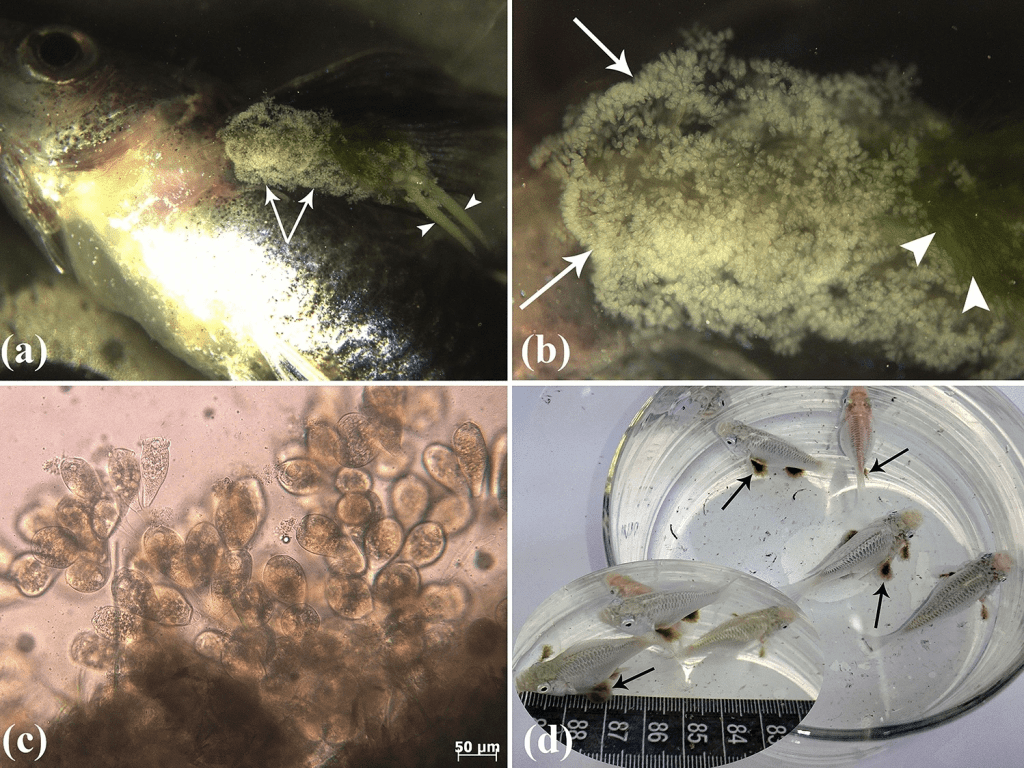
To put it simply, Epistylis is a genus 200 spp. ciliate protozoa that can parasitise on fishes often as a secondary pathogen. It is but appears as a little round ciliated bulb on a stalk, much like Vorticella or rotifers (Wu et al., 2021). In fact though the pathology of the genus in regards to parasitism on fishes has little references to how it looks from the naked eye. Instead the accompanying primary infection e.g. Aeromonas sp. displaying red spots/lesions (Fig 1) but Epistylis niagarae has been suggested to cause scale erosion (Ksepka & Bullard, 2021; Chapman et al., 1976), there is no mention in the literature of Epistylis displaying spots on the body (Pádua et al., 2016).

Instead it seems Epistylis is most accurately identified using a microscope due to it’s distinctive appearance. In Rahmati-Holasoo et al. (2023) it discusses the attachment of Lernaea to the host fish and displays clear images of the common, Epistylis wuhanensis.
It seems that Epistylis is not reported as the rapid killer although the Aerosomas infections could have been the cause, unlike many misconceptions (Chapman et al., 1976).
Is it white spot then?
I can’t say it is, I can say though a microscope should be needed to be sure. I have seen many cysts on fishes and without a microscope it’s just identifying that it’s a lump. Tapeworm cysts I have identified though largely using a microscope but they are larger and do not respond to treatment.
Without a microscope there is a lot of diagnosis going blindly in and even then. There are many resources for identifying fish parasites using microscopy, it’s important though to remember that not all pathogens have been discovered and there are exceptions. Many similar taxa usually can be treated by the same compounds.
Why does it matter?
From my understanding although Epistylis is generally a secondary parasite and not the cause of the infection, it’s opportunistic it usually is susceptible to the same treatments as white spot. Over use of treatments particularly when not targeting the parasite can lead to resistance and I do wonder if that is sometimes what we are seeing. This is more an element of fact checking and should you trust your information sources? They can claim science but do they cite their sources or read theirs? Experience only goes so far but experience doesn’t change what species is what.
References:
Chapman, W. R., Harris, F. A., & Miller, R. W. (1976). Incidence and seasonal variations of Epistylis among fishes in North Carolina reservoirs. In Proceeding of the Annual Conference of Southeastern Association of Game and Fish Commissioners (Vol. 30, pp. 269-275).
Ksepka, S. P., & Bullard, S. A. (2021). Morphology, phylogenetics and pathology of “red sore disease”(coinfection by Epistylis cf. wuhanensis and Aeromonas hydrophila) on sportfishes from reservoirs in the South‐Eastern United States. Journal of Fish Diseases, 44(5), 541-551.
Pádua, S. B. D., Martins, M. L., Valladão, G. M. R., Utz, L., Zara, F. J., Ishikawa, M. M., & Belo, M. A. D. A. (2016). Host-parasite relationship during Epistylis sp.(Ciliophora: Epistylididae) infestation in farmed cichlid and pimelodid fish. Pesquisa Agropecuária Brasileira, 51, 520-526.
Rahmati-Holasoo, H., Marandi, A., Shokrpoor, S., Goodarzi, T., Ziafati Kafi, Z., Ashrafi Tamai, I., & Ebrahimzadeh Mousavi, H. (2023). Clinico-histopathological and phylogenetic analysis of protozoan epibiont Epistylis wuhanensis associated with crustacean parasite Lernaea cyprinacea from ornamental fish in Iran. Scientific Reports, 13(1), 14065.
Rogers, W. A. (1971). Disease in fish due to the protozoan Epistylis (Ciliata: Peritricha) in the southeastern US. In Proc. 25th Ann. Conf. Southeastern Assoc. Game and Fish Comm., 1971 (pp. 493-496).
Wu, T., Li, Y., Zhang, T., Hou, J., Mu, C., Warren, A., & Lu, B. (2021). Morphology and molecular phylogeny of three Epistylis species found in freshwater habitats in China, including the description of E. foissneri n. sp.(Ciliophora, Peritrichia). European Journal of Protistology, 78, 125767.
