Fake news was the word of the year for 2017, we are often bombarded by information and it can be very difficult to fact check. Sometimes we just don’t have time to check the information we are given is correct, and other times we are relying on the credential of the writer. The aquarium hobby is no exception with so many different websites and social media platforms all arguing for space and more importantly your trust.
As distrust in authority increases there has never been so much division and this leads into the influences of certain media. Unlike the general hobby there is a strong influence of science, whether the website is actually scientific or not.
This is best discussed in terms of two of the biggest myths that have arisen in the last few years.
How Epistylis ruled the world.
For many years the protozoan whitespot, Ichthyophthirius multifiliis was widely considered the most common parasite on fishes, it is well studied in the scientific literature (Francis-Floyd et al., 2016, 2023) followed by velvet, Oodinium and Piscinoodiniasis. Disease is tricky, unlike identifying animals we don’t necessary have the tools as a hobby for an accurate diagnosis, there are thousands of species to separate.
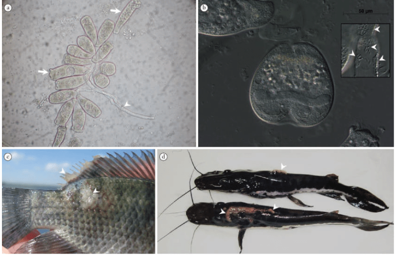
Realistically these two pathogens are one of many that can appear as spots on the fishes body, generally most treatments will cover them. Although there is suggestion that the formalin based treatments are less effective as I discuss here, maybe this has lead to why people come to think they are not dealing with white spot? Because shouldn’t white spot treatments work for I. multifiliis? In addition there is proven acquired immunity to I. multifiliis (Teixeira Alves and Taylor, 2020) but it doesn’t mean that it is unseen. In fact I have noticed a reduced number personally and online of those spotted parasitic cysts, but that is just experience. To add to the confusion not all of these parasites do appear as spots (Sudhagar et al., 2022; Fig 1). I. multifilliis and Piscinoodiniasis is known to target the gills of fishes.
This confusion isn’t aided by nematode cysts looking incredibly similar to I. multifilliis and that used to be quite a common misdiagnosis. Going back to the difficulties in identification, microscopes are very important here as many of these pathogens are really only different to look at under the microscope. Using a microscope is one thing but identifying what you are looking at is another, the koi world seems to have this sorted with pathologists available but it seems to have been a reduced talent within the aquarium hobby. There are very few useful books on pathology still available, most if all will be second hand. Microscopes are really only useful for what can be seen so bacteria and even more viruses are another ball game, requiring expensive technology to identify the pathogen.
So, the story is set for Epistylis. Why Epistylis? We will never know but it is likely a random page that came up and sometimes names stick. Unlike Ichthyophthirius multifiliis, Epistylis is easy to remember. I don’t think the why matters but more that it is there, it’s difficult to work out what website started it.

It might be that fact checking takes time and we should trust what we read to be true. but Ichthyophthirius multifiliis looks nothing alike Epistylis in any way that can be confused as you can clearly see in figure 2 and 3.
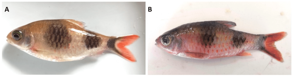
What is more clear is that Epistylis is almost always present and not always as a pathogen, it can also be asymptomatic (Ksepka et al., 2021). But if symptomatic it is expressed as more of a plaque that could be confused with some of the herpes viruses (Fig 3).

So why are they confused? It has to do with this graph (Fig 4). Firstly ich is referring to Ichthyophthirius multifiliis, that we more then often call whitespot in the UK. No citations are provided so this is not backed up with the identification of Epistylis.
So some will say the science is wrong, the issue is that genera are described with set features and if those features don’t match then that is not that species, it would be another. That’s how scientific descriptions work. Epistylis doesn’t display itself as distinct round spots whether rough sized or not (Ksepka et al., 2021; Wu et al., 2021; Valladao et al., 2015). If it was so common and lethal there would be more literature when in reality there isn’t that much comparatively. In my experience as well white spot can kill a fish rapidly, and due to the effect on the gills it is quite taxing on the fish (Martins et al., 2015).
So why this whole essay? To discuss misinformation I must first state why this myth isn’t true. Interestingly it is a brilliant story regarding understanding critical analysis.
How to identify a reliable website:
- Ability to cite their sources, ideally papers if they are scientists.
- Using standard length, just because there are no reliable measurements of total size including the caudal/tail fin.
- Avoiding plagiarism, if a scientist this is a key issue.
References:
Francis-Floyd, R., Yanong, R., & Pouder, D. (2023). Ichthyophthirius multifiliis (white spot) infections in fish.
Francis-Floyd, R., Yanong, R., & Pouder, D. (2016). Ichthyophthirius multifiliis (White Spot) Infections in Fish: CIR920/FA006, rev. 12/2016. EDIS, 2016(10).
Ksepka, S. P., & Bullard, S. A. (2021). Morphology, phylogenetics and pathology of “red sore disease”(coinfection by Epistylis cf. wuhanensis and Aeromonas hydrophila) on sportfishes from reservoirs in the South‐Eastern United States. Journal of Fish Diseases, 44(5), 541-551.
Martins, M. L., Cardoso, L., Marchiori, N., & Benites de Pádua, S. (2015). Protozoan infections in farmed fish from Brazil: diagnosis and pathogenesis. Revista Brasileira de Parasitologia Veterinária, 24, 1-20.
Sudhagar, A., Sundar Raj, N., Mohandas, S. P., Serin, S., Sibi, K. K., Sanil, N. K., & Raja Swaminathan, T. (2022). Outbreak of Parasitic Dinoflagellate Piscinoodinium sp. Infection in an Endangered Fish from India: Arulius Barb (Dawkinsia arulius). Pathogens, 11(11), 1350.
Teixeira Alves, M., & Taylor, N. G. (2020). Models suggest pathogen risks to wild fish can be mitigated by acquired immunity in freshwater aquaculture systems. Scientific Reports, 10(1), 7513.
Valladao, G. M. R., Levy-Pereira, N., Viadanna, P. H. D. O., Gallani, S. U., Farias, T. H. V., & Pilarski, F. (2015). Haematology and histopathology of Nile tilapia parasitised by Epistylis sp., an emerging pathogen in South America. Bulletin of the European Association of Fish Pathologists, 35(1), 14-20.
Wang, Z., Zhou, T., Guo, Q., & Gu, Z. (2017). Description of a new freshwater ciliate Epistylis wuhanensis n. sp.(Ciliophora, Peritrichia) from China, with a focus on phylogenetic relationships within family Epistylididae. Journal of Eukaryotic Microbiology, 64(3), 394-406.
Wu, T., Li, Y., Zhang, T., Hou, J., Mu, C., Warren, A., & Lu, B. (2021). Morphology and molecular phylogeny of three Epistylis species found in freshwater habitats in China, including the description of E. foissneri n. sp.(Ciliophora, Peritrichia). European Journal of Protistology, 78, 125767.
